
The Radon theorem states that the image reconstruction is possible from projections: an image distribution function could be obtained from an infinite set of its projections acquired by the rotational scanning. The algorithms used for CT image reconstruction are based on mathematical foundations of the Radon transformation (Radon theorem) published in 1917 by Johann Radon (Austrian mathematician), who also provided the formula for the inverse transform. The typical energy used in general CT is in the range of 100 kV to 150 kV. CT scanner operations are based on X-rays. The term “computed” in CT (computed tomography) indicates calculated or reconstructed, and the term “tomography” is a compound word comprising the term “tomo” (which meaning to “cut” or “section” in Greek) and “graphy” (which means “to describe” in Greek). Information disappears owing to overlap, tissues with minute absorption coefficients cannot be easily discriminated, and scattering X-rays cause adverse influences on imaging formation. This is generally referred to as radiography. X-rays which penetrate through objects are recorded on films or detectors as two-dimensional images. X-rays are irradiated and penetrate through three-dimensional objects. Röentgen called this radiation type X-ray radiation. This radiation has fluorescence characteristics, sensitizes the film, and penetrates opaque objects. This has been attributed to Wilhelm Conrad Röentgen (Germany) who conducted the first cathode tube experiments (Crookes tube) in Novem. The discovery of X-ray radiation has been a major scientific breakthrough. Keywords : Computed tomography, Physical principle, Clinical application, Technical aspects, Radiation dose The understanding of CT characteristics will provide more effective and accurate patient care in the fields of diagnostics and radiotherapy, and can lead to the improvement of image quality and the optimization of exposure doses. This article reviews the essential physical principles and technical aspects of the CT scanner, including several notable evolutions in CT technology that resulted in the emergence of helical, multidetector, cone beam, portable, dual-energy, and phase-contrast CT, in integrated imaging modalities, such as positron-emission-tomography一CT and single-photon-emission-computed-tomography一CT, and in clinical applications, including image acquisition parameters, CT angiography, image adjustment, versatile image visualizations, volumetric/surface rendering on a computer workstation, radiation treatment planning, and target localization in radiotherapy. Unlike conventional X-ray imaging (general radiography), CT reconstructs cross-sectional anatomical images of the internal structures according to X-ray attenuation coefficients (approximate tissue density) for almost every region in the body. Abstract The evolution of X-ray computed tomography (CT) has been based on the discovery of X-rays, the inception of the Radon transform, and the development of X-ray digital data acquisition systems and computer technology. The toolkit is accessible as a module at. The toolkit is released under the BSD license, imposing minimal restrictions on its use and distribution. Case studies in scan protocol optimization, low dose imaging and iterative algorithm comparison demonstrated its substantial potential in performing scan data based clinical studies. The performance was validated on real patient scans with good agreement with respect to vendor-designed program. Imaging dose in CTDIw is calculated in an empirical formula. Pixel-to-HU mapping is calibrated by a Catphan TM 504 phantom. Reconstruction is supported by TIGRE with FDK as well as a variety of iterative algorithms. Data conditioning operations including scatter correction, normalization, beam-hardening correction, ring removal are performed sequentially.
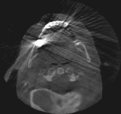
#BEAM HARDENING ARTIFACT MEANING SERIES#
Raw scan data are first decoded to extract X-ray fluoro image series and set up the imaging geometry.

The entire imaging chain, module-based architecture, data flow and techniques used in the creation of the toolkit are presented. The aim of this project is to provide not only a tool to bridge the gap between clinical usage of CBCT scan data and research algorithms but also a framework that breaks down the imaging chain into individual processes so that research effort can be focused on a specific part. We presented TIGRE-VarianCBCT, an open-source toolkit Matlab-GPU for Varian on-board cone-beam CT with particular emphasis to address challenges in raw data preprocessing, artifacts correction, tomographic reconstruction and image post-processing.


 0 kommentar(er)
